Web Content Viewer
Actions
A New Era of Veterinary Care: Harnessing Live X-Ray for Rapid Orthopedic Injury Diagnosis in Patients
Published on 4/11/24
Just like us, our beloved pets are susceptible to orthopedic injuries. Whether it's a playful pup with a sore leg or an aging cat experiencing joint stiffness, understanding the underlying cause of their pain is crucial for effective treatment.
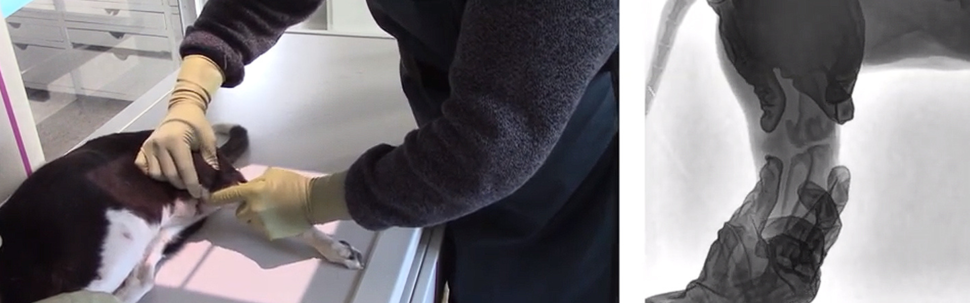
How can veterinary medicine address the challenge of diagnosing complex orthopedic conditions in patients who can’t tell you where it hurts? That's where Fluoroscopy comes in. Fluoroscopy is a real-time X-ray imaging technique that surpasses the static snapshots of standard X-ray images. It employs a continuous stream of low-dose X-rays, generating a live x-ray video on your screen. This enables veterinarians to observe the movement of bones, joints, and even internal organs in real-time, offering a far more dynamic view compared to traditional X-rays.
While Fluoroscopy stands as an excellent diagnostic resource, not every veterinary practice possesses the equipment or expertise to utilize it. However, with recent advancements in veterinary solutions, a comprehensive solution has emerged that is within reach of most veterinary clinics. This innovative technology integrates X-ray capabilities, thermal imaging, patient thickness measurement for automated technique settings, and Fluoroscopy, all into one system.
Let’s delve into the orthopedic injury diagnosis world and discover how fluoroscopy (or as it is sometimes called, live video X-ray) can assist us in performing minimally invasive orthopedic diagnostics and procedures.
Cruciate Ligament Rupture: The Key to a Stable Knee
When a pet presents with lameness or instability in a hind limb, veterinarians can utilize fluoroscopy to assess the health of the cruciate ligament. This minimally invasive technique involves gentle manipulation of the limb under sedation. Fluoroscopy allows the veterinarian to observe any abnormal joint movements indicative of ligament damage, such as cranial drawer or tibial thrust, in real time.
This detailed visualization provides crucial insights into the exact location and severity of the injury, leading to a more precise diagnosis and informed treatment plan. Furthermore, fluoroscopy can be a valuable tool during surgery to repair the ruptured ligament. By offering real-time feedback, it helps veterinarians ensure the accurate placement of implants and confirm joint stability once the repair is complete.
Link to video: https://youtu.be/w6W5usBqaTQ
Complex Mandibular Fractures: Seeing the Bigger Picture
The complex structure of the mandible, or jawbone, makes diagnosing fractures, especially multiple fractures, a challenge with traditional X-rays. Fluoroscopy offers a significant advantage in these cases. By gently manipulating the jaw under fluoroscopy and observing the movement in real-time, veterinarians can gain a much clearer picture of the fracture pattern.
This dynamic visualization allows for precise observation of the displacement and alignment of fractured segments, including any involvement within the jaw joint (intra-articular involvement). Such detailed insights facilitate a more accurate diagnosis and guide the decision-making process for treatment. This may involve conservative management or surgical repair using internal fixation plates.
Link to video: https://youtu.be/uPvQhHO3vzs
Multiple Pelvic Fractures: Unveiling Hidden Instability
X-rays alone can struggle to detect subtle fractures or luxation in the pelvis due to its complex anatomy. This is where fluoroscopy steps in, offering a valuable solution for pet diagnosis. By manipulating the animal's legs and observing the movement on the fluoroscopic screen in real-time, veterinarians can identify even minor fractures that might be missed on a static X-ray.
This dynamic visualization plays a crucial role in determining the most appropriate treatment plan. It allows veterinarians to assess the severity of the fracture pattern, identify any instability, and determine if the fractures require surgical stabilization or if rest and pain management are sufficient.
Link to video: https://youtu.be/tX73VqVgoP8
A Brighter Future for Animal Care
Fluoroscopy is a game-changer in veterinary care, offering real-time visualization of movement that enables veterinarians to diagnose and treat orthopedic injuries with greater precision and effectiveness. This means quicker recovery periods, better treatment results, and, ultimately, a brighter, healthier future for our pets.
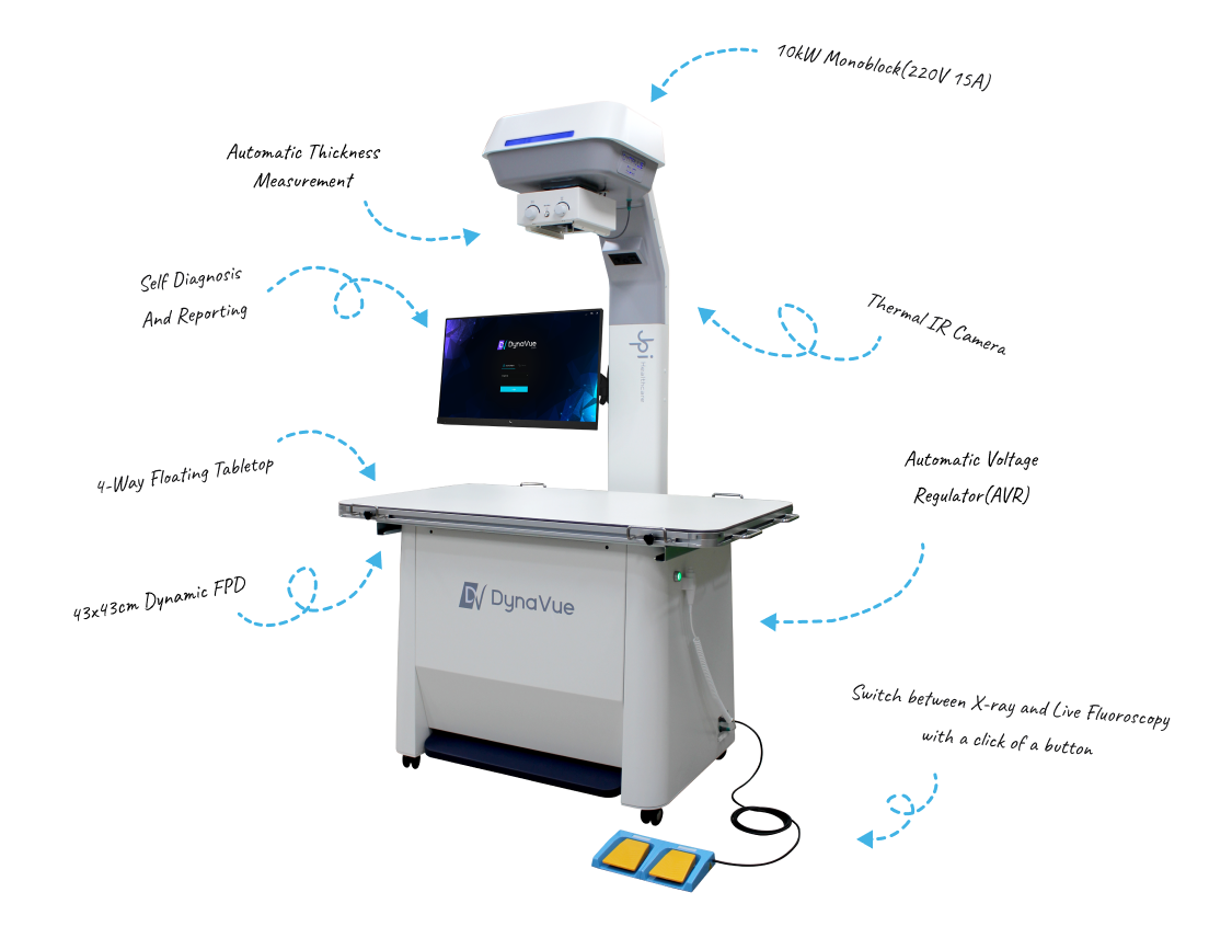
When dealing with an animal with an unidentified injury, the ability to perform X-rays, thermal imaging, or measurements on the patient to pinpoint the precise area requiring diagnosis or conducting a Fluoroscopy without having to move the patient, represents a significant advancement. This is precisely why the DynaVue Duo is revolutionizing veterinary practices.
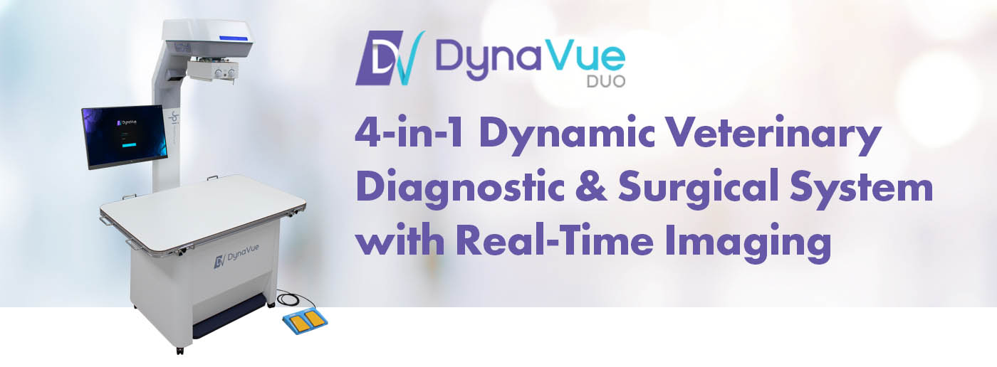
A unique and user-friendly veterinary digital radiography system, DynaVue Duo brings you digital x-ray, fluoroscopy, video, and thermal imaging in a single package for your diagnostic or surgical center.
Designed by the JPI team working with industry-leading veterinarians to optimize workflow, image quality, and diagnostic results. DynaVue Duo is powered by JPI’s ExamVue Duo software, developed specifically with veterinarians' needs and workflow in mind.
Learn More Here